Scintigraphy
Szintigraphie
There are several types of scintigraphy or nuclear medicine imaging tests, including:
- Myocardial scintigraphy (cardiac SPECT) - to evaluate blood flow to the heart muscle and diagnose heart conditions
- Bone scintigraphy (bone scan) - to detect bone cancer, monitor the progression of cancer that has spread to the bones, and evaluate bone injuries or diseases
- Renal scintigraphy (renal scan) - to evaluate the function and blood flow of the kidneys and diagnose kidney conditions
- Lung scintigraphy (ventilation-perfusion scan) - to evaluate lung function and diagnose blood clots in the lungs
- Thyroid scintigraphy - to evaluate the function and structure of the thyroid gland and diagnose thyroid conditions
- Brain scintigraphy (SPECT or PET) - to evaluate brain function and diagnose conditions such as Alzheimer's disease, stroke, and epilepsy
- Granulocyte scintigraphy (Scintimmun)- to detect inflammation or infection in the body
- Gastrointestinal scintigraphy - to evaluate the function and movement of the digestive system and diagnose conditions such as gastroesophageal reflux disease (GERD) and motility disorders.
The specific type of scintigraphy used will depend on the patient's symptoms, medical history, and the condition being evaluated.
The workflow
The workflow for a scintigraphy procedure can vary depending on the type of scintigraphy being performed and the specific protocols of the healthcare facility. However, here is a general overview of the typical steps involved in a scintigraphy procedure:
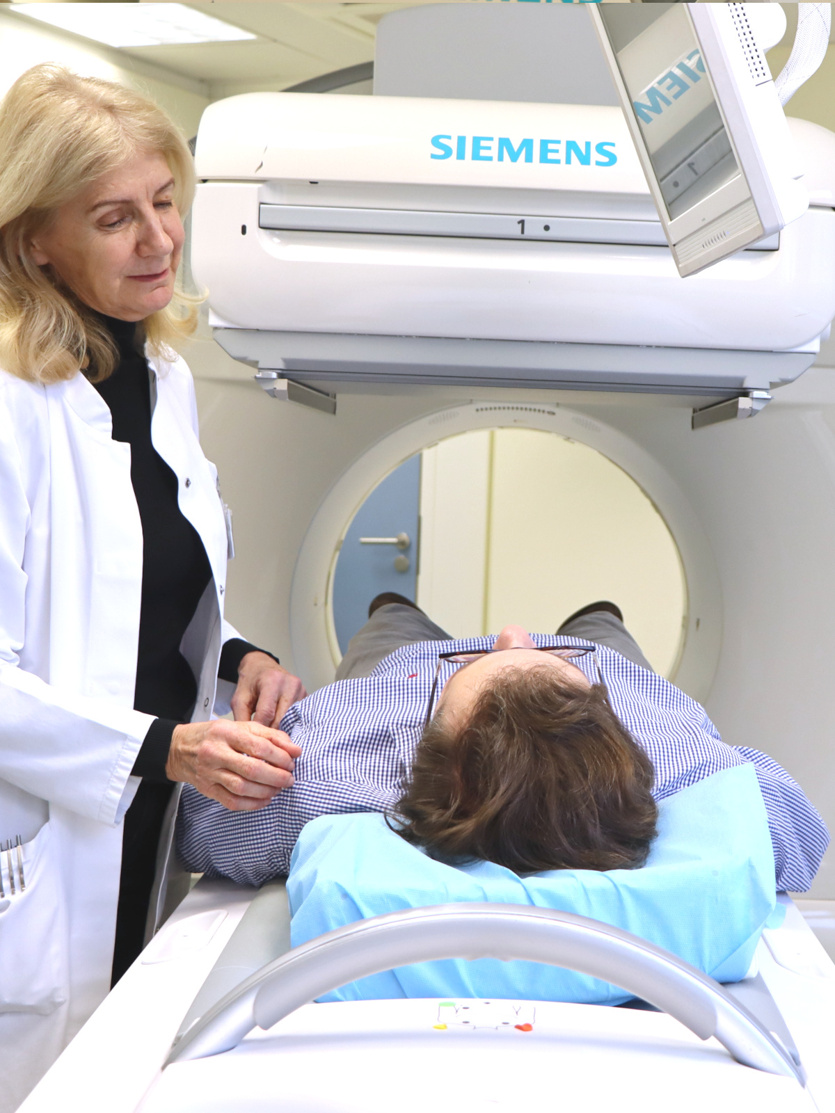
Imaging:
The patient lies on a table and the camera is positioned over the area of interest. The camera takes images of the patient's body, which can take anywhere from a few minutes to several hours depending on the type of scintigraphy being performed.
Preparation:
The patient may be instructed to follow certain dietary or medication restrictions prior to the test. They may also be asked to change into a hospital gown and remove any jewelry or other metal objects.
Injection of radioactive material:
A small amount of radioactive material, called a radiotracer, is injected into the patient's vein, swallowed or inhaled, depending on the type of scintigraphy being performed. The radiotracer travels to the organ or tissue of interest, where it emits gamma rays that can be detected by a special camera.
Imaging:
The patient lies on a table and the camera is positioned over the area of interest. The camera takes images of the patient's body, which can take anywhere from a few minutes to several hours depending on the type of scintigraphy being performed.
Processing of images:
The images are then processed by a computer to create a final image that can be interpreted by a nuclear medicine physician.
After the procedure, the patient can usually resume normal activities, although some types of scintigraphy may require additional precautions, such as avoiding close contact with others for a period of time due to radiation exposure.
The specific instructions and risks associated with the procedure should be discussed with the patient's healthcare provider.